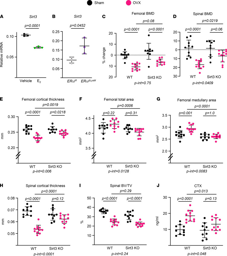
Figure 8. Deletion of Sirt3 attenuates ovariectomy-induced bone loss.
(A and B) Sirt3 mRNA by qPCR in BMMs isolated from 6-month-old female C57BL/6 mice (A) or 3-month-old females of the indicated genotype (B) and cultured with M-CSF (30 ng/mL) and RANKL (30 ng/mL) for 2 days in the presence or absence of E2 (1 × 10⁻⁸M) (triplicate cultures). (C–J) Five-month-old female Sirt3-KO mice and WT littermates were sham operated or ovariectomized (OVX) for 6 weeks (n = 9–11 animals/group). (C and D) Percent change in BMD by DXA 1 day before surgery and before sacrifice. (E–H) Cortical thickness and areas at the femoral midshaft (E–G) and L5 bones measured by micro-CT (H) (n = 9–11 animals/group). (I) BV/TV of trabecular bone in L5 by micro-CT (n = 9–11 animals/group). (J) Serum CTx concentration measured by ELISA (n = 9–11 animals/group). Data are presented as ± SD. P values were determined using Student’s t test (A and B) or 2-way ANOVA (C–J). Interaction terms generated by 2-way ANOVA are shown below each graph. All in vitro assays were performed in cultured BMMs pooled from 3–4 mice/genotype.

