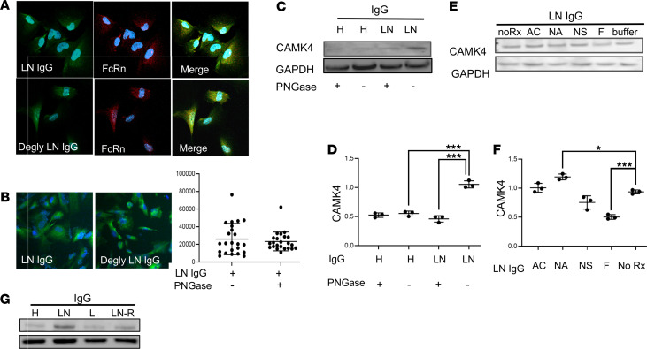
Figure 2. Treatment of IgG with α-fucosidase prevents the upregulation of CaMK4 in podocytes.
(A) Both deglycosylated (Degly) and untreated IgG from individuals with LN colocalize with neonatal Fc receptor (FcRn) in human podocytes (original magnification, 200×). (B) Intensity of Alexa Fluor–tagged PNGase F–treated (deglycosylated) and untreated LN-derived IgG in podocytes was analyzed by immunofluorescence staining (20×). (C) CaMK4 expression was evaluated after exposure of podocytes to deglycosylated or untreated IgG from healthy controls (H) or patients with LN (a representative experiment is shown). (D) Densitometry was performed for quantification of the results in C (3 independent experiments were performed). Data are shown as mean ± SEM. ***P < 0.01, by 1-way ANOVA with Bonferroni post hoc test correction. (E) CaMK4 expression in podocytes after exposure to IgG derived from a patient with LN before (noRx) or after treatment with β-N-acetylglucosaminidase (AC), Neuraminidase A (NA), Neuraminidase S (NS), or α-fucosidase (F). (F) Densitometry was performed for quantification of the results in E. Data are shown as mean ± SEM. *P < 0.05; ***P < 0.01, by 1-way ANOVA with Bonferroni post hoc test correction (3 independent experiments utilizing IgG from 3 different individuals were performed for each representative experiment displayed above). (G) CaMK4 expression was evaluated after exposure of podocytes to IgG differing in glycosylation profiles from individuals with LN in remission, those with SLE with and without nephritis, and those without disease.

