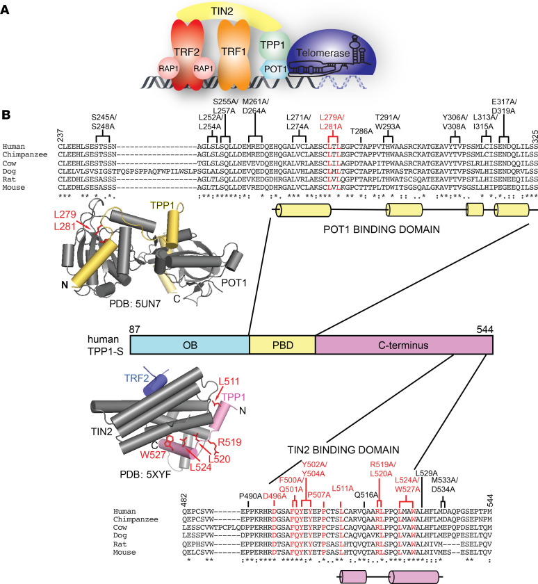
Figure 1. Screen to identify the TPP1 mutations that interfere with POT1 and TIN2 binding.
(A) Schematic representation of the interactions between shelterin proteins and telomerase at chromosome ends. (B) Sequence alignment of the human TPP1 POT1- and TIN2-binding domains with indicated mammalian orthologs. Residues of human TPP1 that were mutated in this screen are shown above the alignment. TPP1 mutants defective in binding POT1 and TIN2 are highlighted in red. Brackets indicate 2 residues simultaneously mutated (double mutant). Asterisks, colons, and periods beneath the sequence lineups represent identical residues, strongly conserved residues, and weakly conserved residues, respectively, as described by the MUSCLE algorithm. Cylinders underneath the sequence alignment indicate α helices. The structure of the POT1 C-terminus bound to the TPP1-PBD (Protein Data Bank [PDB]: 5UN7) is shown above the TPP1 domain diagram with POT1 shown in gray and TPP1 shown in yellow. Structure of the TIN2TRFH-TPP1TBM-TRF2TBM complex (PBD: 5XYF) is shown below the TPP1 domain diagram with TIN2TRFH represented in gray, TRF2TBM represented in purple, and TPP1TBM represented in pink. TPP1 amino acids whose mutation resulted in POT1- and TIN2-binding defects are shown in red in the structures.

