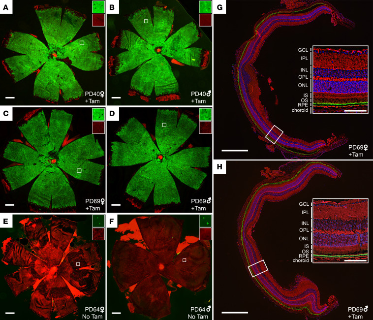
Figure 2. Cre recombinase activity in Rpe65CreERT2 mouse retina assessed with the mT/mG reporter.
(A and B) RPE flatmounts from representative Cre+/– female and male mice, respectively, that were administered IP tamoxifen for 5 consecutive days starting on PD21 (n = 4). The flatmounts were obtained 2 weeks after completion of the tamoxifen regimen and were imaged with a fluorescence microscope. Green fluorescence indicates cells where Cre-mediated recombination has occurred, whereas red fluorescence indicates un-recombined cells. (C and D) RPE flatmounts from representative Cre+/– female and male mice that were administered IP tamoxifen for 5 consecutive days starting on PD50 (n = 7). The flatmounts were obtained 2 weeks after completion of the induction regimen. (E and F) show representative RPE flatmounts from Cre+/– female and male mice, respectively, that were not treated with tamoxifen (n = 6). The flatmounts were obtained on PD64. Scale bars indicate 500 μm. The insets in A–F show zoomed areas (marked by white squares, original magnification, 10×) with green and red channels separately displayed to demonstrate the single-cell resolution of the imaging method. (G and H) Retina cryosections from Cre+/– female and male mice, respectively, that were administered IP tamoxifen for 5 consecutive days starting on PD21 (n = 2). The cryosections were obtained 2 weeks after completion of the induction regimen. Insets show zoomed views of the areas marked with white rectangles with the retinal cell layers labeled. The green label represents Cre-mediated recombination and is restricted to the RPE. The scale bars in G and H indicate 500 μm while those in the insets indicate 100 μm. GCL, ganglion cell layer; INL, inner nuclear layer; IPL, inner plexiform layer; IS, inner segment; ONL, outer nuclear layer; OPL, outer plexiform layer; OS, outer segment; RPE, retinal pigment epithelium; Tam, tamoxifen.

