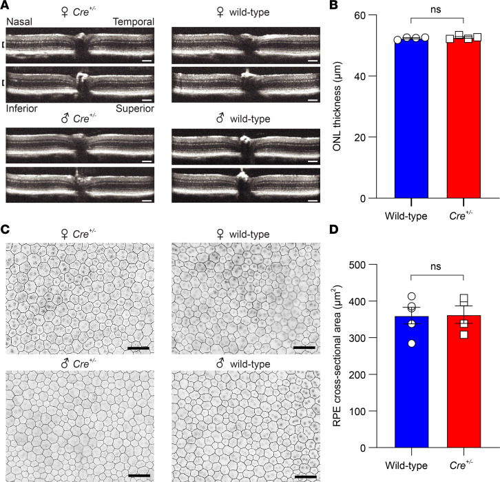
Figure 5. Impact of Cre induction on retinal and RPE structure in Rpe65CreERT2 mice.
(A) Retinal OCT images from representative 4-month-old mice (M450 wild-type Rpe65) that had been treated with IP tamoxifen starting on PD21. The anatomical labeling in the upper left applies to all panels, and the scale bar indicates 100 μm. The laminar retinal structure is indistinguishable between wild-type and Cre+/– mice. (B) Quantification of the ONL thickness (demarcated by black brackets in A) showed no significant difference between wild-type (n = 4) and Cre+/– (n = 4) mice (52.4 ± 0.7 μm vs. 52.6 ± 0.7; P > 0.99, 2-tailed t test). (C) RPE flatmount images from the same mice as in A stained with an anti–ZO-1 antibody to allow visualization of the plasma membrane. The images were acquired within 1000 μm of the optic nerve. The scale bar indicates 50 μm. (D) Quantification of the RPE cross-sectional area showed no significant difference between wild-type (777 measured cells, n = 4) and Cre+/– (742 measured cells, n = 4) mice (360.1 ± 22.5 vs. 362.8 ± 23.8 μm2, respectively; P = 0.94, 2-tailed t test). Each point represents data from a single mouse. Bars indicate means ± SEM.

