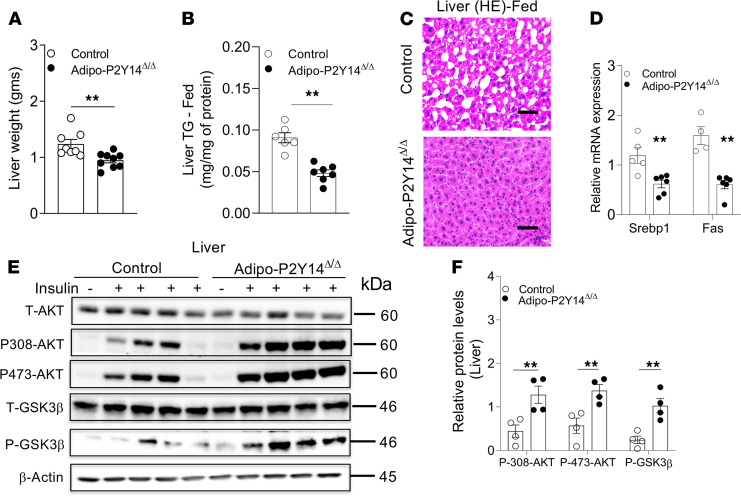
Figure 5. Adipo-P2Y14Δ/Δ mice show protection from liver steatosis and improved hepatic insulin sensitivity.
(A) Liver weight (grams) from adipo-P2Y14Δ/Δ and control mice (n = 8 or 9/group). (B) Liver triglyceride (TG) levels from adipo-P2Y14Δ/Δ and control mice (n = 6–7/group). (C) Representative H&E-stained sections of liver from HFD adipo-P2Y14Δ/Δ and control mice. (D) Gene expression levels of Srebp1 and Fas in liver of HFD adipo-P2Y14Δ/Δ and control mice (n = 4–6/group). (E) Western blot analysis of insulin signaling in liver of HFD adipo-P2Y14Δ/Δ and control mice (n = 4/group). (F) Quantification of immunoblotting data for E (n = 4/group). The expression of 18s rRNA was used to normalize qRT-PCR data. All data are expressed as mean ± SEM. **P < 0.01 (2-tailed Student’s t test). All experiments were conducted on mice consuming an HFD for at least 8 weeks. Scale bar: 150 µm. P2Y, purinergic; HFD, high-fat diet.

