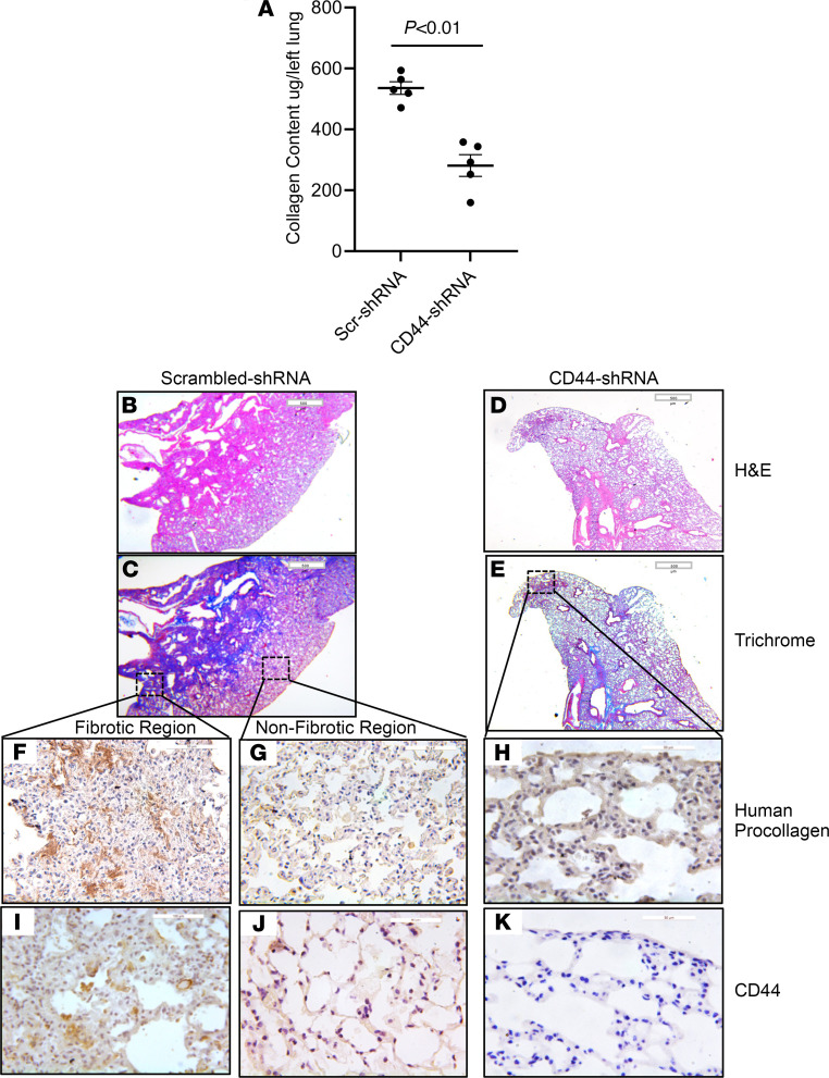
Figure 3. CD44 regulates the fibrogenicity of CD44hi IPF MPCs.
NSG mice were treated with IT bleomycin (1.25 U/kg). Two weeks later, the mice received IPF MPCs (IPF424) transduced with either CD44 shRNA or scrambled shRNA via tail vein injection (106 cells/100 μL); 10 mice/group. Lungs were harvested 4 weeks after cell administration. (A) Collagen content was quantified in left lungs by Sircol assay. (B–K) Serial 4 μm sections of right lung tissue from mice receiving CD44hi IPF MPCs transduced with scrambled-shRNA or CD44-shRNA (B–E scale bar: 500 μm; F, G, I, and J scale bar: 50 μm; H and K scale bar: 20 μm). Representative H&E and Trichrome stains assessing fibrosis and collagen deposition, respectively (B–E). IHC using an antibody-recognizing human procollagen to identify human cells and assess collagen synthesis (F–H) and a CD44 antibody to determine the distribution of CD44-expressing cells (I–K). (F, G, I, and J) IHC for human procollagen (F and G) and CD44 (I and J) to assess the distribution of human cells expressing collagen and CD44-expressing cells from fibrotic and nonfibrotic lung regions from mice receiving CD44hi IPF MPCs transduced with scrambled shRNA. Data are expressed as mean ± SEM. P values in A were determined by 2-tailed t test.

