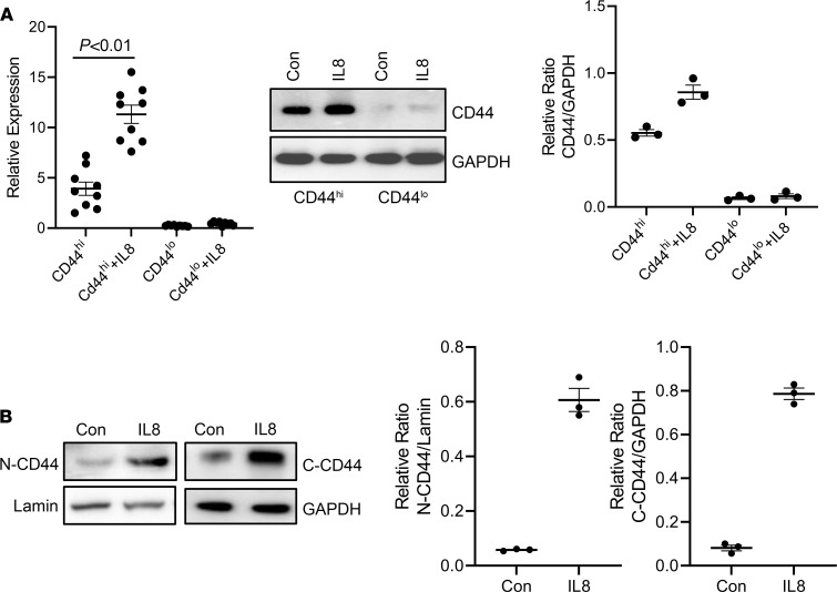
Figure 6. IL-8 increases CD44 expression and nuclear accumulation.
(A) CD44hi and lo IPF MPCs were treated with recombinant IL-8 (5 ng/mL). CD44 mRNA (left panel) and protein (middle panel) expression were quantified by qPCR and Western blot analysis, respectively. Densitometry values summarizing Western blot data in right panel. GAPDH served as loading control. (B) CD44hi IPF MPCs were treated with recombinant IL-8 (5 ng/mL). CD44 protein levels were quantified in nuclear (N) and cytoplasmic (C) fractions by Western blot analysis. Lamin (nuclear) and GAPDH (cytoplasmic) served as loading controls. Densitometry values summarizing Western blot data shown in right graphs. IPF 422, IPF424, and IPF 442 were used in these figures. Data are expressed as mean ± SEM. n ≥ 3 independent experiments for each experimental condition or group except the Western blot is from a single experiment representative of 3 independent replicates. Data are expressed as mean ± SEM. P value in A was determined by 2-tailed Student’s t test.

