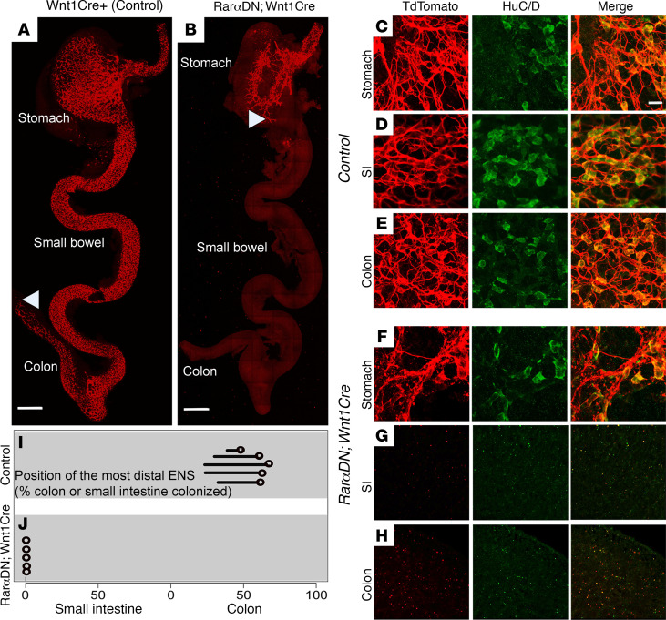
Figure 2. Cell-autonomous RAR signaling is required for ENS precursor colonization of fetal small bowel and colon.
(A and B) Representative images of bowel colonization by TuJ1+ enteric neurons (red) at E12.5. (A) Wnt1Cre+ controls have TuJ1+ cells throughout the bowel from esophagus to midcolon. (B) RarαDNLoxP/+; Wnt1Cre+ mice only have TuJ1+ cells present in the esophagus and stomach. A white arrowhead shows the position of the most distal TuJ1+ cell or neurite in each image. Scale bars: 500 μm. (C–H) Representative E12.5 images of stomach, small intestine (SI), and colon show cells stained with TdTomato+ (red) and HuC/D (green) throughout the bowel in control (C–E), whereas TdTomato+- and HuC/D-stained cells are absent in SI and colon of RarαDNLoxP/+; Wnt1Cre+ mice (F–H). (C–H) Scale bar: 150 μm. (I and J) Circles show the position of the most distal TdTomato+ ENS cell in control or RarαDNLoxP/+; Wnt1Cre+ mice at E12.5. The line attached to each circle indicates a hypoganglionic zone in controls. n = 5 controls, n = 5 RarαDNLoxP/+; Wnt1Cre+.

