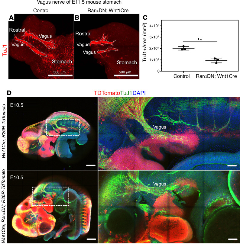
Figure 6. Vagal nerve fibers occupy a smaller area in E11.5 stomach of RarαDNLoxP/+; Wnt1Cre+ mice than in controls.
(A and B) TuJ1 antibody–stained E11.5 stomach of RarαDNLoxP/+ (control) (A) or RarαDNLoxP/+; Wnt1Cre+ (B) mice. Scale bar: 500 μm. (C) Quantification of TuJ1+ stained vagal fiber area. **P < 0.01 by 2 tailed unpaired Student’s t tests. (D) Whole embryo imaging of E10.5 Wnt1Cre+; R26R-TdTomato+ and RarαDNLoxP/+; Wnt1Cre+; R26R-TdTomato+. Scale bars: 500 μm (left) and 200 μm (right, enlarged). Note that TuJ1+ vagal nerve fibers are not TdTomato+. Box shows region of the magnified image. n = 3, Wnt1Cre+; R26R-TdTomato+. n = 3, RarαDNLoxP/+; Wnt1Cre+; R26R-TdTomato+. Supplemental Video 1 and Supplemental Video 2 show 3-dimensional Z-stacks from embryos in D.

