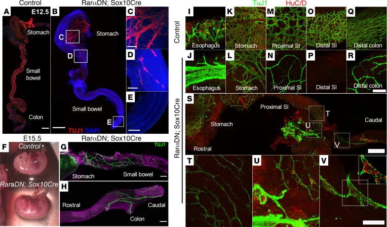
Figure 7. RarαDNLoxP/+; Sox10Cre+ mice had extensive distal bowel aganglionosis.
(A–E) E12.5 control or RarαDNLoxP/+; Sox10Cre+ whole bowel was stained with TuJ1 antibody (red) and DAPI (blue). Insets highlight mutant stomach (C), proximal small intestine (D), and distal small intestine (E). Sparse TuJ1+ nerve cell bodies were seen in the proximal small bowel (D) but not in more distal small bowel (E) of RarαDNLoxP/+; Sox10Cre+ mice. Scale bars: 500 μm (A and B) and 100 μm (C–E). (F) Images of either RarαDNLoxP/+ (control) or RarαDNLoxP/+; Sox10Cre+ E15.5 embryos. Note the defective eye and craniofacial development of the mutant embryos. (G and H) E15.5 RarαDNLoxP/+; Sox10Cre+ bowels were stained with TuJ1 (green) and counter-stained with DAPI. Note the TuJ1+ network at proximal small intestine (G) and distal colon (H). Scale bars: 100 μm. (I–R) RarαDNLoxP/+ (control) or RarαDNLoxP/+; Sox10Cre+ bowels were stained with HuC/D (red) and TuJ1 (green) antibodies. Representative images of esophagus (I and J), stomach (K and L), proximal small intestine (M and N), distal small intestine (O and P), and end of distal colon (Q and R). Scale bar: 50 μm. (S) Proximal small intestine of E15.5 RarαDNLoxP/+; Sox10Cre+ bowel. Regions outlined by boxes indicate which areas are enlarged in (T–V). Note extrinsic nerve fibers (T and U). Scale bars: 500 μm (S) and 50 μm (T–V). n = 3, RarαDNLoxP/+ (control); n = 3, RarαDNLoxP/+; Sox10Cre+.

