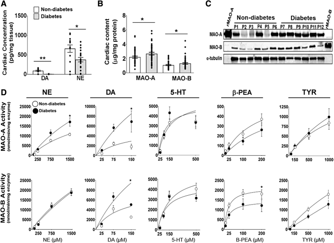
FIG. 1.
Cardiac MAO protein expression and enzymatic activity in human atrial myocardium. LC-MS analysis of DA and NE concentrations in human atrial myocardium (A) from patients with type 2 diabetes and nondiabetes (n = 15/metabolic group). MAO-A and MAO-B concentrations measured by using MAO isoform specific antibodies via (B) ELISA (n = 45–55/metabolic group) and (C) Western blot, α-tubulin was used as the loading control (n = 6/metabolic group). Kinetic traces of MAO activity (D) using 5 biogenic substrates: NE, DA, serotonin, βPEA, and TYR. MAO activity was measured by monitoring the increase in fluorescence of resorufin at Ex/Em: 571/585. MAO-A activity is shown on the top panel and MAO-B activity is on the bottom panel (n = 20/metabolic group). Data are represented as ± SEM. (A, B, D) was analyzed by using unpaired t-test. *p < 0.05, **p < 0.005 compared with nondiabetes. 5-HT, serotonin; βPEA, β-phenylethylamine; DA, dopamine; ELISA, enzyme-linked immunosorbent assay; LC-MS, liquid chromatography–mass spectrometry; MAO, monoamine oxidase; NE, norepinephrine; SEM, standard error of the mean; TYR, tyramine.

