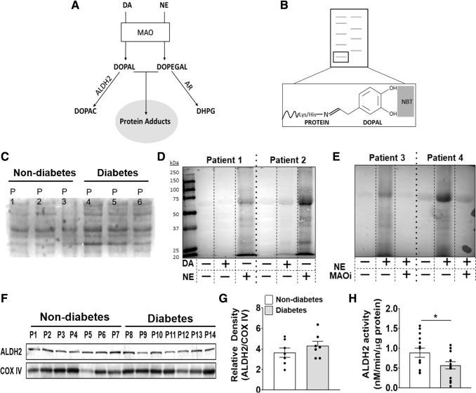
FIG. 4.
Detection and metabolism of catecholaldehydes in human atrial myocardium. Pathway of catecholamine metabolism into catecholaldehydes (by MAO) and then into acetic acid (DOPAC) or alcohol (DHPG) by ALDH2 or AR, respectively (A). If metabolism by ALDH2 or AR is compromised, catecholaldehydes can form carbonyl adducts with proteins. Shown in (B) is a representative image of catechol-protein adduct detection by NBT staining. In the presence of catechol-modified proteins, NBT stains proteins blue on nitrocellulose membrane. Representative NBT stains of cardiac lysate untreated (C), after overnight incubation with DA (25 μM) or NE (75 μM) (D), and overnight incubation with MAO inhibitors ± NE (75 μM) (E). Western blot analysis (F) and densitometric analysis (G) of ALDH2 in myocardium. COX IV was used as a loading control to calculate relative density. n = 7/metabolic group. ALDH2 activity (H) measured by monitoring the increase in absorbance of NADH at 450 nm in myocardial samples. n = 14/metabolic group. (H) Was analyzed by using unpaired t-test. *p < 0.05. All data are shown as mean ± SEM. ALDH2, aldehyde dehydrogenase 2; AR, aldose reductase; COX IV, complex IV; DHPG, dihydroxyphenylglycol; DOPAC, dihydroxyphenylacetic acid; MAOi, monoamine oxidase inhibitors; NBT, nitroblue tetrazolium.

