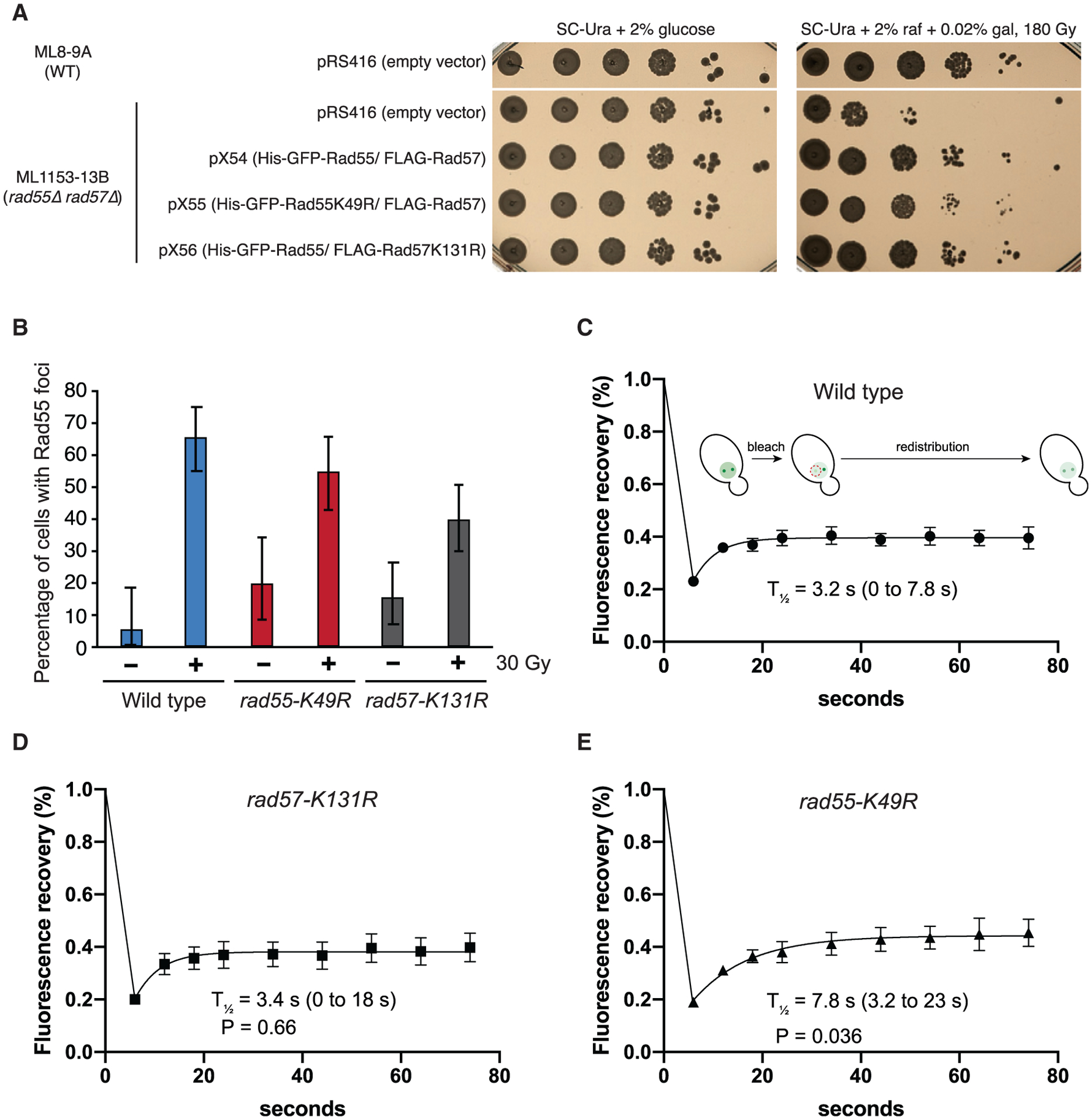
Figure 5. ATP hydrolysis by Rad55 regulates turnover of Rad55-Rad57 in vivo.

(A) WT or rad55Δ rad57Δ cells were complemented with empty vector (pRS416) or vectors expressing His-GFP-Rad55 and FLAG-Rad57 transgenes as shown (pX54, pX55, and pX56). Ten-fold serial dilutions of the indicated strains were plated and subjected to 180 Gy of ionizing radiation (IR).
(B) Cells were exposed to 30 Gy of IR and inspected for Rad55 foci after 2-h incubation at 25°C (n = 37–97). RAD55 and RAD57 transgenes were expressed from plasmids pX54, pX55, or pX56 in rad55Δ rad57Δ cells (ML1153–13B). These conditions yielded an average of two Rad55 foci per cell.
(C–E) Cells were exposed to 30 Gy of IR and subjected to iFRAP, where one Rad55 focus is photobleached, and fluorescence redistribution from the other focus is monitored by time-lapse microscopy (n = 6–9). 95% CIs are indicated in parentheses. Error bars indicate SEM. Data were derived from two biological replicates. The p values are indicated for comparison with the wild type in (C) (Student’s t test).
