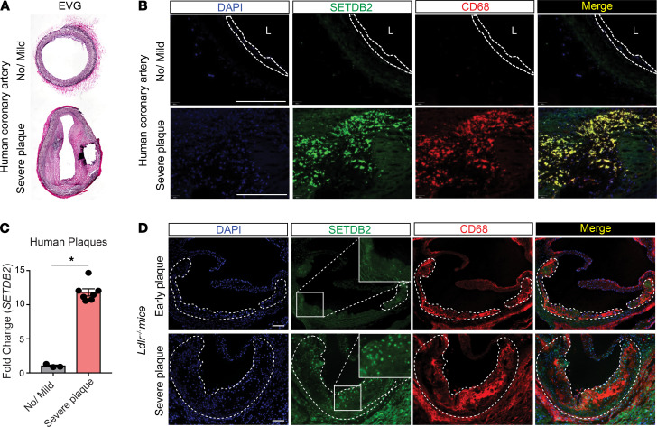
Figure 2. SETDB2 is highly expressed in atherosclerotic macrophages.
(A) Representative elastic van Gieson (EvG) staining from healthy (top) and atherosclerotic (bottom) human coronary arteries. (B) Representative immunofluorescence analysis of SETDB2 and CD68 expression in human coronary lesions. (C) qRT-PCR analysis of Setdb2 expression in healthy (no/mild, n = 3) and atherosclerotic arteries (severe plaque, n = 7). Quantification represents the mean ± SEM of relative expression levels normalized to healthy artery. *P < 0.05. Data were analyzed by an unpaired 2-tailed Student’s t test. (D) Representative immunofluorescence analysis of SETDB2 and CD68 expression in mouse atherosclerotic plaques. Dashed bars indicate the atherosclerotic plaques. Scale bar: 100 μm. L, lumen.

