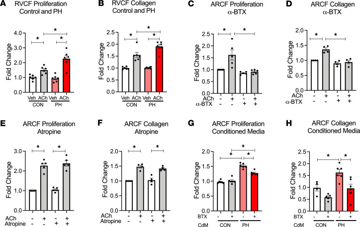
Figure 4. RV cardiomyocyte–derived ACh promotes cardiac fibroblast proliferation and collagen synthesis through α7 nAChR activation.
(A and B) RVCFs isolated from 7-wk control and PH rats treated with vehicle or 10 nM ACh for 24 hours and then assessed for cell proliferation by cell counts (A) and collagen content (B) with Sircol assay (n = 6). (C and D) Cell counts (n = 5) (C) and collagen content (n = 4) (D) of ARCF in response to 10 nM ACh with or without α7 nAChR antagonist α-BTX (100 nM) for 24 hours. (E and F) Cell counts (n = 5) (E) and collagen content (n = 4) (F) of ARCF in response to 10 nM ACh in the presence or absence of muscarinic receptor antagonist atropine (50 μM) for 24 hours. (G and H) Cell count (G) and collagen content (H) from ARCFs treated with conditioned media from isolated 7 wks Con/PH RV cardiomyocytes. Data are shown as mean ± SEM. ANOVA followed by Bonferroni comparison. *P < 0.05. ARCF, Adult rat cardiac fibroblasts; RVCF, right ventricular cardiac fibroblasts; ACh, acetylcholine; α-BTX, α-bungarotoxin; RVCM, right ventricular cardiac myocytes; CdM, conditioned media (from RV cardiomyocytes); CON, control; PH, pulmonary hypertension.

