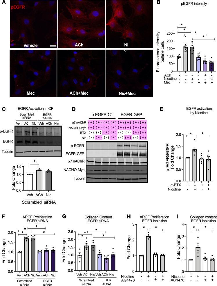
Figure 5. Cardiac fibroblast proliferation and collagen synthesis are mediated via α7 nAChR–EGFR transactivation.
(A) Representative fluorescence image of containing p-EGFR (Y1068) (red) and nuclei (blue by Hoechst) in ARCF 30 minutes after vehicle, ACh (10 nM), or nicotine (600 nM) treatment in the presence or absence of Mec (20 μM). Scale bar: 20 μm. (B) Summary data from A (n = 9). (C) EGFR transactivation in ARCFs treated with vehicle, 600 nM nicotine, or 10 nM ACh for 30 minutes. EGFR activation was estimated by p-EGFR(Y1068)/EGFR (n = 3). EGFR siRNA and scrambled siRNA-transfected cells were used to confirm the antibody specificities. (D) EGFR transactivation in HEK293T-overexpressing EGFR, α7 nAChR, and Myc-tagged NACHO, by nicotine (600 nM, 30 min) in the presence or absence of α-BTX. Cells stably overexpressing EGFP were used as a control. EGFR phosphorylation, overexpressed EGFR, α7 nAChR, and NACHO were detected by the antibodies against p-EGFR (Y1068), GFP, α7 nAChR, and Myc, respectively. (E) Summary data from D (n = 6). p-EGFR/EGFR was normalized to that in cells without nicotine and α-BTX treatment. (F and G) Cell counts (n = 4) (F) and collagen content (n = 4) (G) of ARCF in response to ACh (10 nM) or nicotine (600 nM) in cells transfected with either EGFR specific or scrambled siRNA. (H and I) Cell counts (n = 5) (H) and collagen content (n = 5) (I) of ARCF in response to nicotine (600 nM) in presence or absence of EGFR inhibitor AG1478 (50 μM). Data are shown as mean ± SEM. ANOVA followed by Bonferroni comparison. *P < 0.05. ACh, acetylcholine; α-BTX, α-bungarotoxin; AG1478, EGFR inhibitor; Mec, mecamylamine; nAChR, nicotinic acetylcholine receptor.

