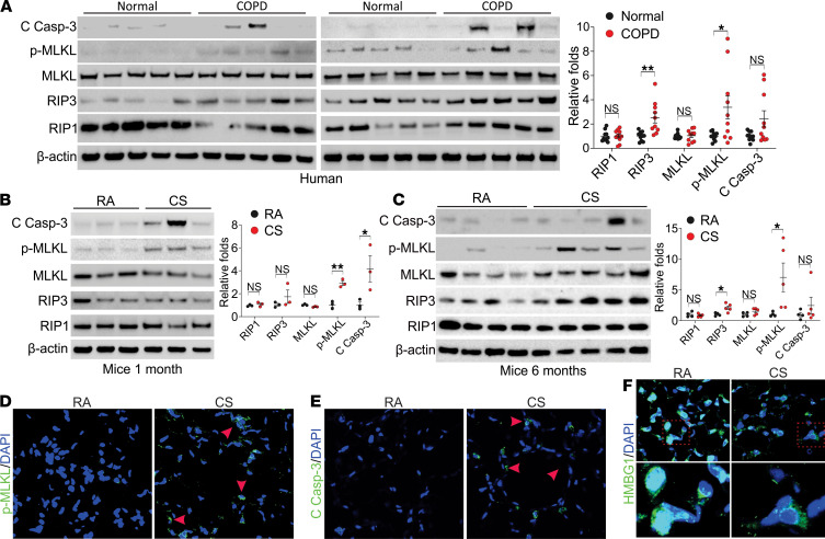
Figure 1. Necroptosis and apoptosis were detected in the lung tissue from COPD patients and CS treated mice.
(A) Western blot analysis of cleaved caspase-3 (C Casp-3), p-MLKL (S358, p-MLKL), MLKL, RIP3, and RIP1 in the lung tissue from donors and patients with COPD. Left, the representative pictures; right, the quantification of each protein’s expression. (B and C) Western blot analysis of C Casp-3, p-MLKL (S345, p-MLKL), MLKL, RIP3, and RIP1 in the lung tissue from C57BL/6J mice exposed to room air (RA) or CS for 1 month (B) and 6 months (C). Left, the representative pictures; right, the quantification of each protein’s expression. (D–F) Immunofluorescence staining of p-MLKL (D), C Casp-3 (E), and HMGB1 (F) in lung tissue from mice exposed to RA or CS for 6 months. Each experiment was repeated 3 times. Original magnification, ×400 (D and E), ×600 (F). Red arrowheads indicate the immunofluorescence-positive cells. Data represent the means ± SEM. *, P < 0.05; **, P < 0.01. Student’s t test was conducted for each 2-group comparison.

