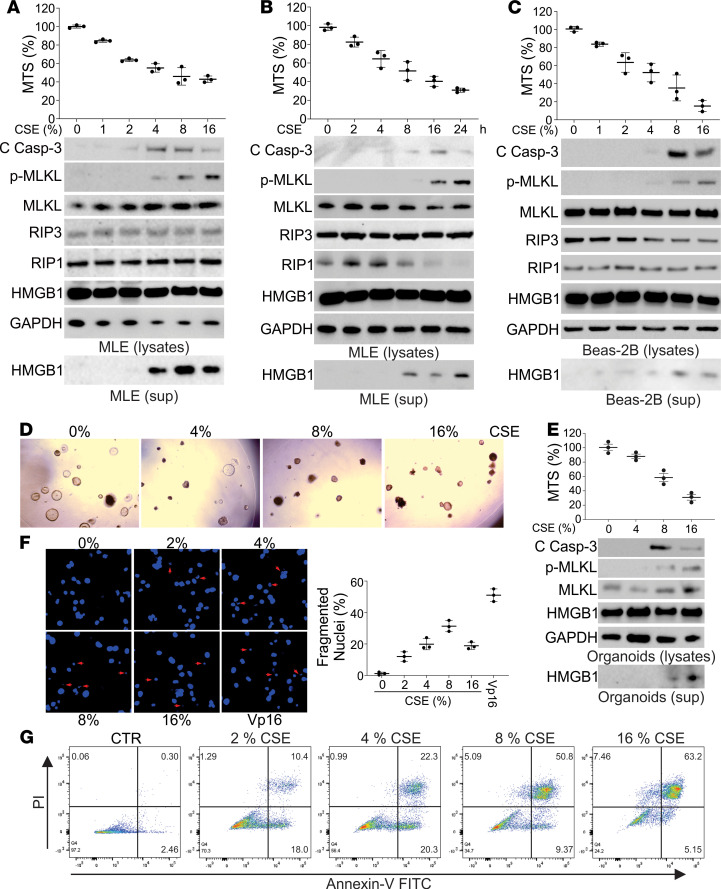
Figure 2. Necroptosis and apoptosis were detected in the CSE-treated lung epithelial cells and organoids.
(A) MLE-12 cells were treated with the indicated concentration of CSE for 16 hours. Upper, the cell viability was analyzed by MTS assay; lower, the expression of indicated proteins in cell lysates and HMGB1 in cell supernatant was analyzed by Western blot. (B) MLE-12 cells were treated with 8% CSE for the indicated time. Upper, the cell viability was analyzed by MTS assay; lower, the expression of indicated proteins in cell lysates and HMGB1 in cell supernatant was analyzed by Western blot. (C) Beas-2B cells were treated with the indicated concentration of CSE for 16 hours. Upper, the cell viability was analyzed by MTS assay; lower, the expression of indicated proteins in cell lysates and HMGB1 in cell supernatant was analyzed by Western blot. (D) The organoids derived from mouse lungs were treated with indicated concentrations of CSE for 16 hours. The representative pictures of organoids were shown. (E) The organoids were treated as in D. Upper, the cell viability was analyzed by MTS assay; lower, the expression of indicated proteins in cell lysates and HMGB1 in cell supernatant was analyzed by Western blot. (F) The nuclei fragmentation of MLE-12 cells treated with the indicated concentration of CSE for 16 hours was analyzed using Hoechst 33258 staining. Etoposide (Vp-16) (25 μM) was used as a positive control. Left, the representative pictures; right, the percentage of fragmented nuclei was counted. Original magnification, ×600. Arrow indicates the fragmented nuclei. (G) The apoptosis and necroptosis of MLE-12 cells treated with indicated concentrations of CSE were analyzed by annexin V/PI staining followed by flow cytometry analysis. The representative data were shown. Data represent the means ± SEM. Each experiment was repeated 3 times. PI, propidium iodide.

