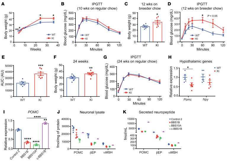
Figure 6. BBS1M390R mutation reduces POMC expression in both mouse hypothalamus and human iPSC–derived hypothalamic neurons.
(A) Body weight curve of male WT and BBS1M390R-knockin (KI) mice (n = 9, 10). (B) Intraperitoneal glucose tolerance test (IPGTT) of 10-week-old male mice on regular chow diet (n = 9, 10). (C and D) Body weight (C) and IPGTT (D) of 12-week-old male mice fed breeder chow ad libitum (n = 6, 7). (E) The glucose area under the curve (AUC) in WT and KI mice as shown in D. ***P < 0.001. (F) Body weight of 24-week-old WT and KI mice on regular chow diet (n = 9, 8). (G) IPGTT of 24-week-old WT and KI mice on regular chow diet (n = 9, 8). (H) qPCR analysis of Pomc and Npy expression in hypothalamus of WT and KI mice (24-week-old males) after 16-hour fasting followed by 4-hour refeeding (n = 5, 5). *P < 0.05, **P < 0.01 by 2-tailed Student’s t test (A–H). (I) qPCR analysis of POMC expression in day 35 iPSC-derived hypothalamic neurons (n = 3). **P < 0.01, ****P < 0.0001 by 1-way ANOVA followed by Tukey’s multiple-comparison test. (J and K) Amount of neuropeptide produced in neuronal lysates (J) and in cultured medium (16-hour culture, K) from control (n = 3), BBS1B (n = 3), BBS10A (n = 3), and c-BBS1B (n = 1) iPSC–derived hypothalamic neurons. POMC, αMSH, and βEP concentrations were measured with ELISA and radioimmunoassay and normalized to total protein.

