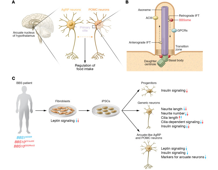
Figure 1. Bardet-Biedl syndrome proteins control primary cilia function in hypothalamic neurons.
(A) In the brain, the arcuate nucleus of the hypothalamus is central for controlling energy homeostasis in the body. Here, the interplay of proopiomelanocortin (POMC) and Agouti-related protein (AgRP) neurons, which require primary cilia for their normal function, regulates food intake. (B) Primary cilia originate from the basal body, a modified mother centriole, which is part of the centrosome. The basal body serves as a nucleation site for microtubules, which form the core structure of all cilia, the axoneme. Along the axoneme, the intraflagellar transport (IFT) machinery transports proteins anterogradely to the ciliary tip or retrogradely to the ciliary base via the molecular motors kinesin-2 and dynein-2, respectively. The transition zone at the ciliary base prevents free diffusion of proteins from the cell body into the cilium. The BBSome is a central component, which, together with the IFT, regulates the protein content in the cilium. (C) Wang et al. (14) differentiated generic neurons as well as POMC and AgRP neurons from BBS and non-BBS patient–derived iPSCs (via fibroblasts). Using this human in vitro system, the authors uncovered the effect of mutations in BBSome-encoding genes on neuronal differentiation and on the central signaling pathways regulating the function of POMC and AgRP neurons in energy homeostasis. Arrows indicate decrease or increase. Note that some aspects were only investigated in cells with BBS1 (blue arrows) or BBS10 (red arrows) mutations.

