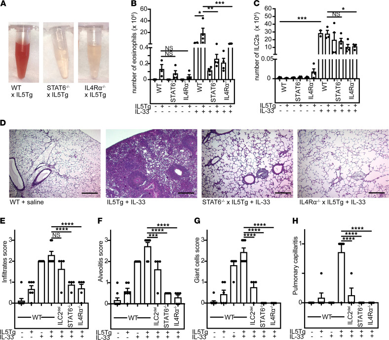
Figure 4. Deficiency of IL4Rα signaling protects from vasculitis in hypereosinophilic mice.
(A) BAL from IL-33–treated IL5Tg, STAT6–/– x IL5Tg, or IL4Rα–/– x IL5Tg. Representative of 4 or more mice/group. (B) Eosinophils in BAL of mice from A. Data are presented as ± SEM. ****P < 0.0001; **P < 0.01; *P < 0.05; ns = by 1-way ANOVA with Sidak post hoc testing. n = 2–4 mice/group. (C) ILC2s in BAL of mice from A. ***P < 0.001; *P < 0.05; ns = by 1-way ANOVA with Sidak post hoc testing. n = 2–4 mice/group. (D) H&E staining of lungs from representative IL-33–treated IL5Tg, STAT6–/– x IL5Tg or IL4Rα–/– x IL5Tg mice treated with IL-33, as shown in A–C (original magnification, ×10). Scale bars: 100 μm. (E) Histopathological scoring for infiltrates, (F) alveolitis, (G) giant cells, and (H) pulmonary capillaritis in ILC2del, STAT6–/–, or IL4Rα–/– with or without IL5Tg after saline or IL-33 treatment. ****P < 0.0001, ***P < 0.001, **P < 0.01; *P < 0.05 by 1-way ANOVA with Dunnett’s post hoc testing. n = 4–9 mice/group. Data are presented as ± SEM. BAL, bronchoalveolar lavage; ILC2s, type 2 innate lymphoid cells.

