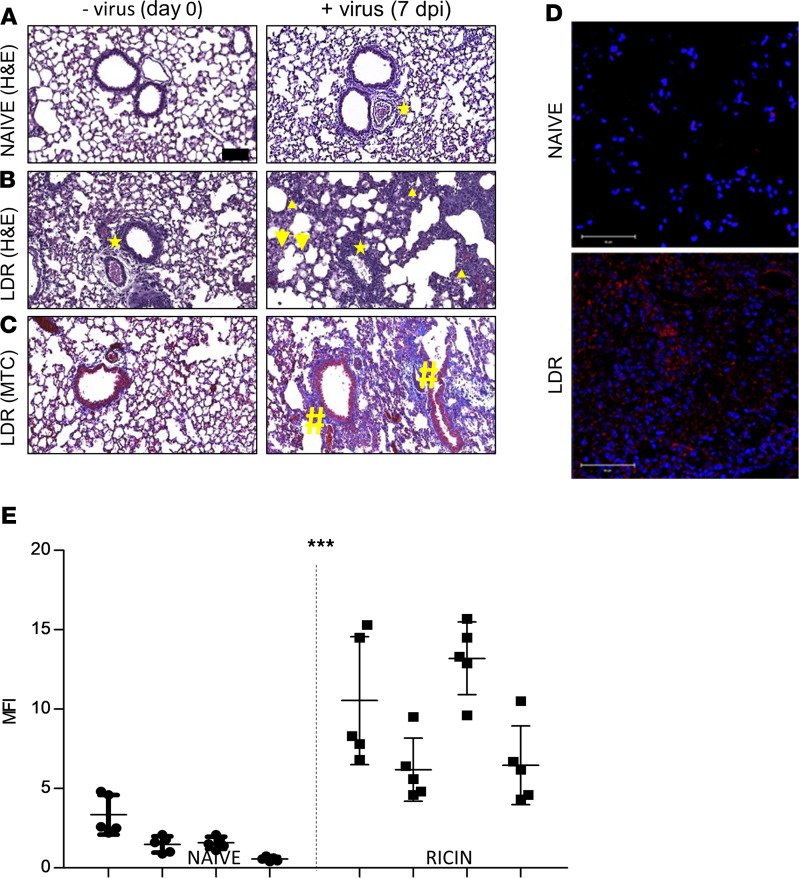
Figure 4. SARS-CoV-2 binding and injury in lungs of LDR mice.
(A–C) Histology: naive (A) and LDR (B and C) mice (ricin application at day –2) were subjected to SARS-CoV-2 infection at a dose of 5 × 10⁶ PFU/mouse. Lungs at the time of viral infection (day 0) and 7 days later were harvested, fixed, and processed for paraffin embedding. Sections (5 μm) were stained with H&E for general histopathology (A and B) or with Masson’s trichrome (MTC) for collagen (C). Representative sections of n = 4 mice are shown. Indicated are perivascular and peribronchial inflammation (stars), infiltration in perivascular and alveolar sites (triangles), edema (arrows), and fibrin (hashtags). Magnification ×20, scale bar: 100 μm. (D and E) Confocal microscopy scans of lung sections. (D) Sections from LDR mice (lungs harvested 2 days after LDR administration) and naive mice were incubated with SARS-CoV-2 (1000 PFU/mL), immunostained with a mAb directed against the spike region of SARS-CoV-2, and then visualized by AF594-labeled anti-human antibody. Staining of SARS-CoV-2 in red and identification of nuclei by DAPI in blue, scale bar: 50 μm (see also Supplemental Figure 3). Representative sections of n = 4 mice are shown. (E) Scatterplots of the fluorescence staining intensities of SARS-CoV-2 expressed as MFI ± SEM, n = 4 mice, 5 scanned fields/lung, analyzed using unpaired t test. ***P <0.001. Abbreviation: dpi, days after infection.

