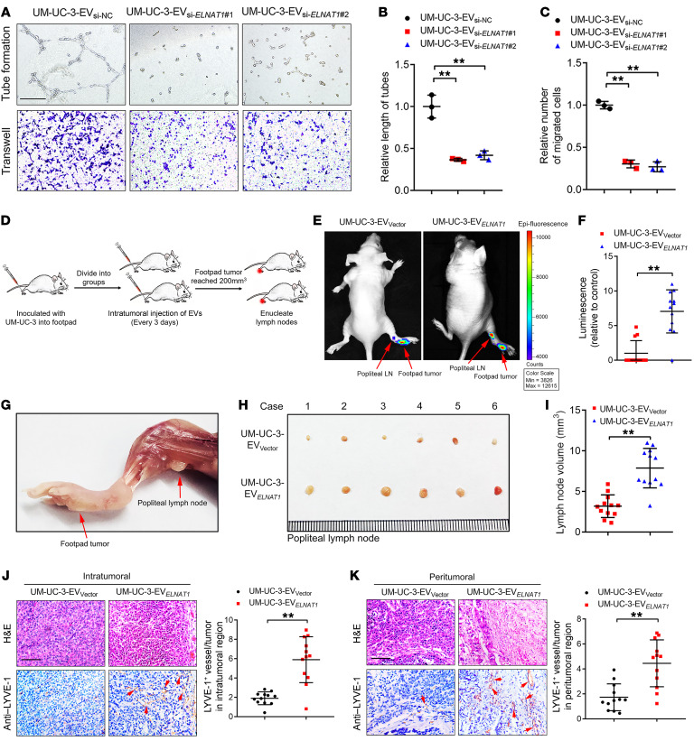
Figure 3. EV-mediated ELNAT1 facilitates lymphangiogenesis and lymphatic metastasis of BCa in vitro and in vivo.
(A–C) Representative images (A) and quantification of tube formation (B) and Transwell migration (C) of HLECs treated with UM-UC-3-EVsi-NC, UM-UC-3-EVsi-ELNAT1#1, or UM-UC-3-EVsi-ELNAT1#2. Scale bar: 100 μm. A 1-way ANOVA followed by Dunnett’s test was used to assess statistical significance. (D) Schematic representation for establishing the nude mouse model of popliteal LN metastasis. (E and F) Representative bioluminescence images and quantification of metastatic popliteal LNs from nude mice in the UM-UC-3-EVVector and UM-UC-3-EVELNAT1 groups (n = 12). The red arrows indicate the footpad tumor and metastatic popliteal LN. A 2-tailed Student’s t test was used to assess statistical significance. (G–I) Representative images of popliteal LNs and quantification of the LN volume for the UM-UC-3-EVVector and UM-UC-3-EVELNAT1 groups (n = 12). A 2-tailed Student’s t test was used to assess statistical significance. (J and K) Representative IHC images and quantification of lymphatic vessels in the intratumoral and peritumoral regions of footpad tumors (n = 12). Scale bars: 50 μm. A 2-tailed Student’s t test was used to determine statistical significance. Error bars show the SD of 3 independent experiments. **P < 0.01.

