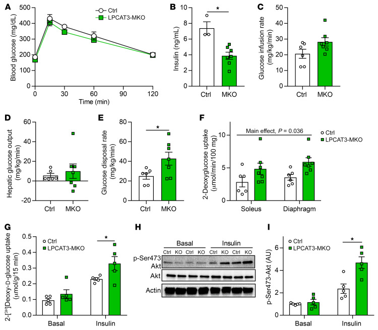
Figure 6. LPCAT3-MKO mice are protected from diet-induced skeletal muscle insulin resistance.
(A) Intraperitoneal glucose tolerance test (Ctrl n = 6, MKO n = 8). (B) Serum insulin at the 30-minute time point of the glucose tolerance test (Ctrl n = 3, MKO n = 8). (C–F) Hyperinsulinemic-euglycemic clamps were performed in conscious unrestrained mice (Ctrl n = 6, MKO n = 7). (C) Glucose infusion rate required to maintain constant blood glucose of 150 mg/dL during clamp phase. (D) Hepatic glucose output during the clamp phase. (E) Rate of whole-body glucose disposal during the clamp phase. (F) 14C-2-deoxyglucose uptake quantification in soleus and diaphragm muscles during the clamped state. (G–I) Soleus muscles were dissected and incubated with or without 200 μU/mL of insulin. (G) Ex vivo 2-deoxyglucose uptake (n = 5). (H and I) Ser473 phosphorylation and total Akt (n = 5). All data are from HFD-fed mice. Two-way ANOVA with Šidák’s multiple-comparison test (A, F, G, and I) or 2-tailed t tests (B–E) were performed. All data are represented as mean ± SEM. *P ≤ 0.05.

