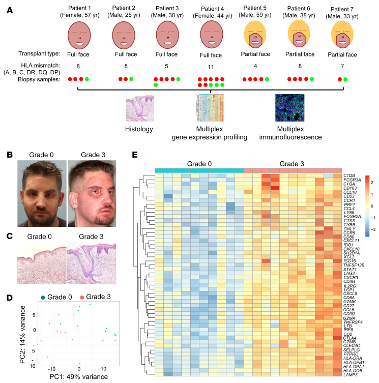
Figure 1. Human face transplant rejection has a distinct gene expression signature.
(A) Design of the study. Skin biopsies from 7 face transplant patients collected during episodes of acute cellular rejection (red) and nonrejection (green) were analyzed using histologic examination, multiplex gene expression profiling, and immunostaining. (B) Clinical photographs of a recipient of a full face transplant during nonrejection (grade 0) and severe acute cellular rejection (grade 3), demonstrating edema and erythema of the transplanted face. (C) Representative examples of H&E staining of a face transplant skin biopsy graded as nonrejection (grade 0, minimal inflammatory infiltrates), and a second biopsy graded as severe acute cellular rejection (grade 3, dermal inflammatory infiltrates with apoptotic keratinocytes). (D) Unsupervised principal component analysis clustered grade 3 rejection biopsies (n = 11) separately from grade 0 samples (n = 10). (E) Heatmap of the top 50 genes differentially expressed in grade 3 compared with grade 0 biopsies (log2 fold change >1; adjusted P value <0.05). Differentially expressed genes (DEGs) were obtained using normalized gene expression counts as input and the Wald significance test. Each column represents a facial allograft biopsy. Gene values are row scaled. The full list of differentially expressed genes and associated statistics are shown in Supplemental Table 3.

