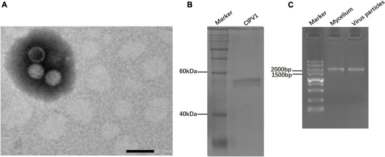
FIGURE 5.
Virus particles isolated from the mycelia of C. liriopes strain Cl-B-2. (A) Isometric, non-enveloped viral particles with diameter of 35 nm were observed by transmission electron microscope (TEM). (B) SDS-PAGE electrophoresis (12%) analysis of the purified virus particles showing the protein band of coat protein. (C) Agarose gel electrophoresis of dsRNA extracted from the mycelia and viral particles, respectively.

