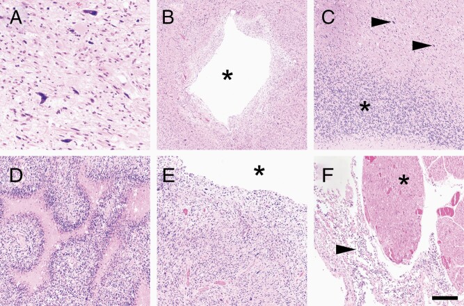
Figure 2.
Postmortem findings. (A) Postmortem examination of the original tumor resection cavity showed abundant viable tumor cells, including markedly atypical cells, as was seen prior to immunotherapy. Granulomas, eosinophils, and plasma cells were no longer present anywhere in the brain. Tumor spread diffusely throughout all brain regions sampled, including the midbrain (B, asterisk = central aqueduct), cerebellum (C, asterisk = granular neurons, arrows = tumor cells), pons (D), and medulla (E, asterisk = fourth ventricle). Leptomeningeal dissemination was present, all the way down into the spinal cord, around the nerve rootlets in the thoracic region (F, arrow = tumor, asterisk = nerve rootlet). Scale bar in (F) = 80 μm in (A), 400 μm in (B), 200 μm in (C–F).

