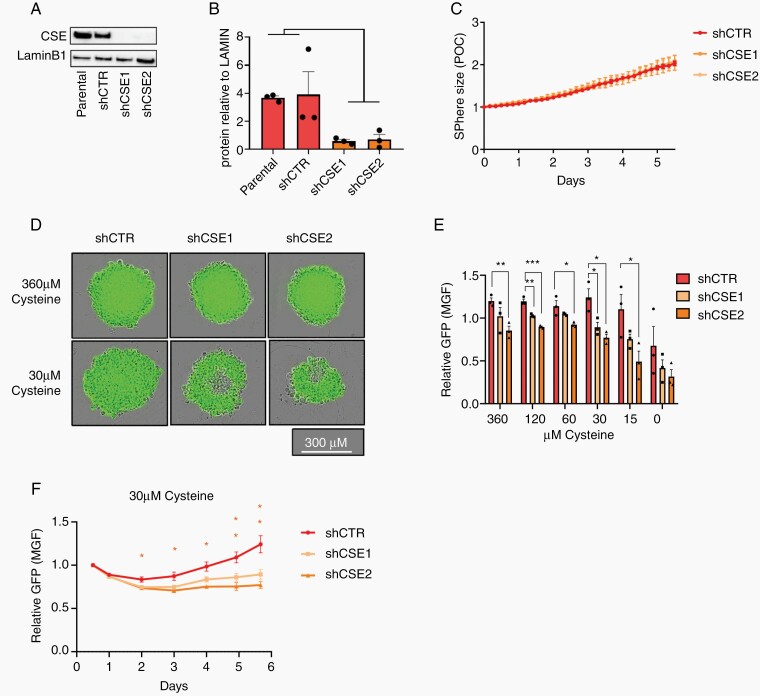
Figure 2.
CSE is required to maintain viability of IDH1m astrocytoma cells under cysteine depletion. (A) CSE western blot analysis and (B) quantification relative to LaminB1 in IDH1m AS cells (NCH1681) with 2 CSE KD clones (shCSE1, shCSE2) compared to the parental and scramble control (shCTR) (n = 3). (C) Relative sphere size under standard culture conditions (360 µM cysteine). Data presented as fold change of phase object confluence (POC) of each cell line relative to day 0 (n = 3 with 5 spheres per experiment). (D) Representative fluorescent images of spheres after 6 days. (E) Relative GFP signal in CSE KD cells at decreasing cysteine concentrations (day 6). Measurements expressed as fold change of mean green fluorescence (MGF) of each cell line relative to starting point (360 µM cysteine at day 0.5 once the sphere was formed) (n = 3 with 5 spheres per experiment). (F) Time course of GFP signal of spheres at 30 µM cysteine. All data presented as means ± SEM. * P < .05, ** P < .01; *** P < .001.

