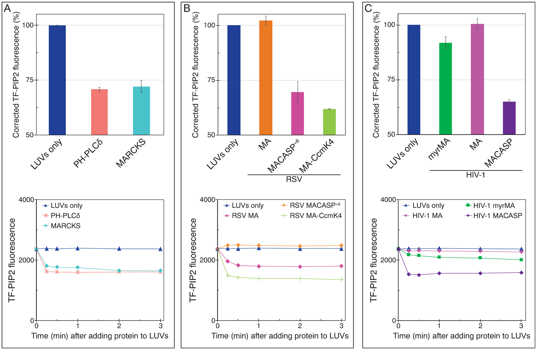
Figure 5.

HIV-1 and RSV Gag-derivative proteins can induce PIP2 clustering, as measured by fluorescence quenching. LUVs with 2% PIP2 were prepared as in Figure 2 using 1 mM EDTA to eliminate all multivalent cation binding. Protein at a final concentration of 20 μM was added after LUV formation, and thus the inner leaflet of the liposomes was not exposed to protein. Top panels: TF-PIP2 fluorescence quenching by (a) control proteins, (b) RSV membrane binding proteins, and (c) HIV-1 membrane binding proteins. TF-PIP2 fluorescence of LUVs mixed with buffer was set to 100%, and the effect of added protein was recorded. Note that for protein-induced PIP2 clustering, the Y-axis is shown only from 50% to 100%, since TF-PIP2 fluorescence quenching occurred only from the outer leaflet where lipids were exposed to the proteins. Each bar in the graph represents the average TF-PIP2 fluorescence % from three time points 1, 2, and 3 min post-mixing. Each quenching assay was performed at least three times with error bars of standard deviations from the means. (a–c, bottom panels) Time course of quenching. For each assay, TF-PIP2 fluorescence was measured at 0.25, 0.5, 1, 2, and 3 min post-mixing.
