FIG 1.
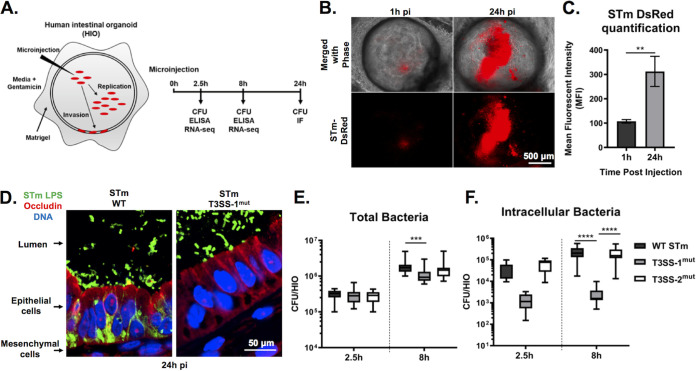
WT S. Typhimurium (STm) replicates within the lumen of HIOs and invades IECs dependent on T3SS-1. (A) Diagram of experimental protocol. (B) Fluorescence microscopy of HIOs injected with S. Typhimurium-DsRed, a strain that harbors the pGEN plasmid encoding red fluorescence protein (DsRed) (10). (C) Quantification of panel B. n = 3 biological replicates. Error bars represent SD. P = 0.0047 by unpaired t test. (D) Immunofluorescence of HIO sections infected with S. Typhimurium WT (left) and S. Typhimurium T3SS-1mut (right). LPS, lipopolysaccharide. (E) Total bacteria in HIOs at 2.5 and 24 h postinjection. n = 16 biological replicates. Whiskers represent minimum and maximum values. Significance was calculated by two-way analysis of variance (ANOVA). (F) Intracellular bacteria in HIOs at 24 h postinjection. n > 31 biological replicates. Whiskers represent minimum and maximum values. Significance was calculated by Mann-Whitney test.

