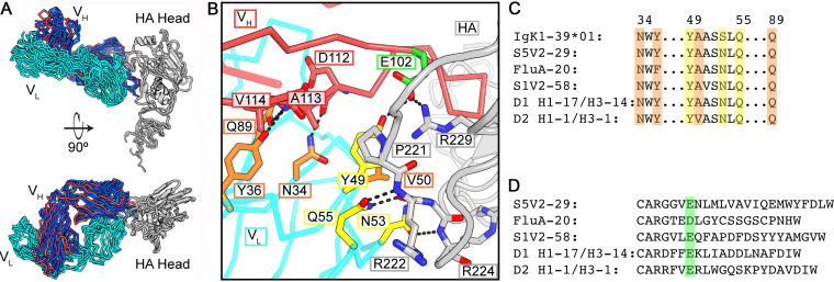
FIG 2.
Stereotyped HA engagement by IGκV1-39 antibodies. (A) Superposition on the HA head domain of the five IGκV1-39 antibodies. The HA heads are in gray, IGκV1-39 is in cyan, and heavy chains are colored according to VH gene usage and are the same as those in Fig. 1. (B) Zoomed-in view of the IGκV1-39 interaction of D2 H1-1/H3-1 with HA from the bottom view of panel A. Key residues are shown in sticks. Light chain residues that contact HA are in yellow, and those that buttress/permit a curled HCDR3 conformation are in orange. A common acidic residue in HCDR3 is highlighted in green. (C) Partial sequence alignment of the germ line IGκV1-39 sequence and the five antibodies. Shading is colored as described above for panel B. (D) Sequence alignment of the antibody HCDR3s. The acidic residue at the sixth position of HCDR3 is shaded in green.

