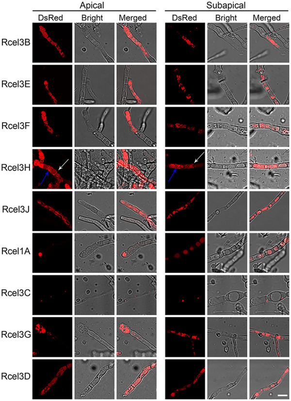
FIG 5.

Cellular localizations of CEL3B-DsRed, CEL3E-DsRed, CEL3F-DsRed, CEL3H-DsRed, CEL3J-DsRed, CEL1A-DsRed, CEL3C-DsRed, CEL3G-DsRed, and CEL3D-DsRed at the apical and subapical regions of nine recombinant strains, Rcel3B, Rcel3E, Rcel3F, Rcel3H, Rcel3J, Rcel1A, Rcel3C, Rcel3G, and Rcel3D, respectively. The confocal images were observed under CLSM at 120 h for all strains except Rcel3D, which was monitored at 168 h. The white and blue arrows indicate the septa and organelle membranes of Rcel3H, respectively. All the strains were grown in TMM plus 2% cellulose. Scale bar = 10 μm.
