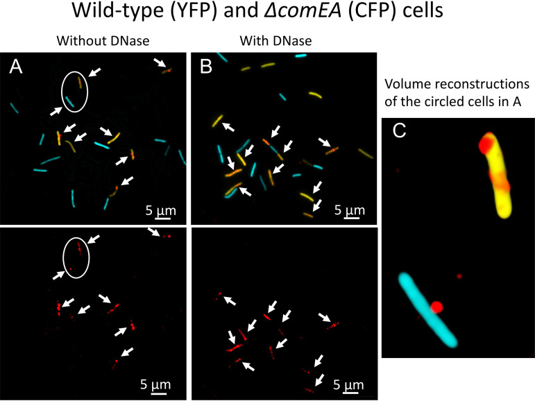
FIG 4.
Binding and uptake in the ΔcomEA mutant. Wild-type (YFP) and ΔcomEA cells were combined before incubation for 30 min with rDNA. Panel A shows the cells without DNase treatment, and panel B shows cells with treatment. In the top images, the rhodamine, CFP, and YFP channels were merged, and the bottom images in panels A and B show only the rhodamine channel. The arrows indicate the positions of all the rhodamine signals. Panel C shows one aspect from a 3D deconvolution of the CFP- and YFP-expressing cells circled in panel A.

