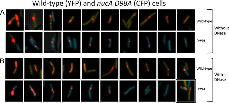
FIG 5.
Binding and uptake in the nucA D98A mutant. (A and B) Wild-type (YFP) and D98A (CFP) mutant cells were made competent, combined, and after 15-min incubation with rDNA, imaged without (A) and with DNase (pB). In each of these panels, the top and bottom rows show nearly all the wild-type and mutant cells with rDNA signals from a single field. The rhodamine signal was enhanced before cropping the cells, and the intensities of the rhodamine signal can be directly compared within the panels. The YFP and CFP images were separately adjusted in each image so as not to obscure the rDNA signal. The images in these two panels were ordered by apparent rDNA signal strengths, decreasing from left to right, to facilitate comparisons of the mutant and wild-type cells. The boxed image in panel B shows a cell expressing both YFP and CFP.

