FIG 1.
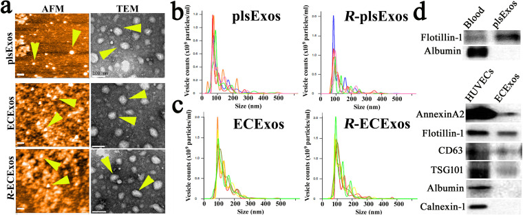
Characterization of plsExos and ECExos after SEC isolation. (a) plsExos and ECExos morphologies were verified using atomic force microscopy (AFM) (left; scale bars, 200 nm) and transmission electronic microscopy (TEM) (right; scale bars, 100 nm). (b and c) The vesicle size distribution of isolated EVs was analyzed using nanoparticle tracking analysis (NTA) (n = 5 per group). (d) Expressions of indicated protein markers in 100 μg of proteins of plsExos (upper portion) and ECExos (lower portion) were examined using Western immunoblotting.

