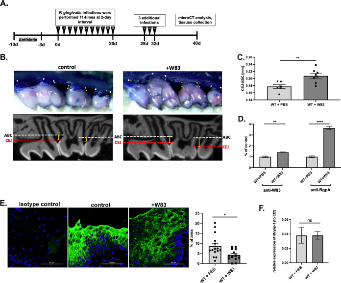
FIG 1.
Repeated infection with P. gingivalis W83 induces bone loss in a murine model, contributing to depletion of MCPIP-1 protein in gingiva. (A) Graphical representation of P. gingivalis-induced periodontitis in mice. (B) Visualization of P. gingivalis W83-induced bone loss in wild-type (WT) mice. Representative images of methylene blue (upper) and sectional micro-computed tomography (micro-CT) analysis of gingival tissue (bottom) with indicated cementoenamel junction (CEJ) to alveolar bone crest (ABC) distances on the second molar (yellow lines). (C) Quantitative analysis of the CEJ-ABC distances of the second molar. n = 6 in control group; n = 8 in infected group (+W83). (D) Levels of anti-W83 and anti-RgpA IgG antibodies in murine sera. Results for the control mice are shown as 1. (E) Visualization of MCPIP-1 protein level (green) by confocal laser scanning microscopy in murine gingiva, along with protein quantification presented as a percentage of area of the fluorescence signal. Analysis performed based on 15 images. (F) Relative expression of Mcpip-1 transcript in gingival tissue. All data represent mean values ± standard error of the mean (SEM). *, P < 0.05; **, P < 0.01; ****, P < 0.0001; ns, not significant.

