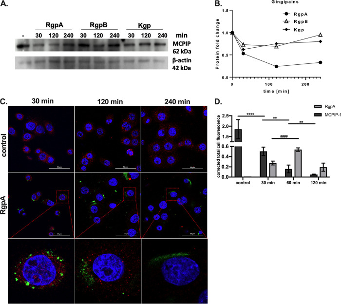
FIG 3.
RgpA degrades MCPIP-1 protein in gingival keratinocytes. (A) Representative Western blot analysis, using specific anti-MCPIP-1 antibodies, of protein lysates from TIGK cells treated with 2 nM gingipains (RgpA, RgpB, or Kgp) for the indicated times. (B) Densitometry analysis demonstrating MCPIP-1 protein fold change normalized to β-actin level in TIGK lysates. (C) Visualization of MCPIP-1 protein (red) and RgpA (green) level by confocal laser scanning microscopy in TIGKs. (D) Quantitative analysis of MCPIP-1 and RgpA fluorescence signal. Data show mean fluorescence values (± standard deviation [SD]) in 8 tested fields of view. **, P < 0.01; ****, P < 0.0001; ####, P < 0.0001.

