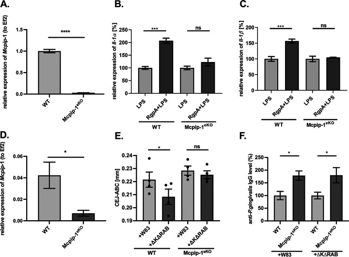
FIG 5.
The LPS sensitivity of keratinocytes depends on MCPIP-1 expression. (A) Depletion of Mcpip-1 in keratinocytes determined by qRT-PCR. (B, C) Wild-type (WT) and Mcpip-1-deficient (Mcpip-1eKO) murine keratinocytes were exposed to RgpA (2 nM) for 1 h, followed by stimulation with LPS (20 μg/ml) for an additional 3 h. Relative expression of (B) IL-1α and (C) IL-1β mRNA was determined by qRT-PCR. Cells stimulated with LPS are presented as 100%. (D) Relative expression of Mcpip-1 transcript in murine gingival tissue (n = 4). (E) Quantitative analysis of the CEJ-ABC distances of the second molar of the P. gingivalis-induced bone loss in WT and Mcpip-1eKO mice after repetitive infections with wild-type strain W83 or with the gingipain-deficient ΔKΔRAB mutant (n = 4 in each group). (F) Levels of anti-P. gingivalis IgG antibodies in murine sera. The values in WT individuals were defined as 100%. All data represent mean values ± SEM. *, P < 0.05; **, P < 0.01; ***, P < 0.001; ****, P < 0.0001; ns, not significant.

