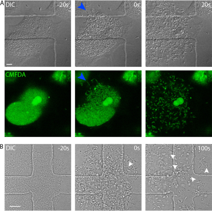
FIG 2.

T. cruzi egress is a rapid event. (A) Vero cells, infected with T. cruzi trypomastigote forms, were visualized by confocal live-cell imaging up to parasite egress. −20 s, pre-egress; 0 s, moment of egress; 20 s, after egress. Images were acquired with a capture interval of 20 s. Cells were preincubated with CMFDA. Blue arrowheads indicate the site of membrane rupture. The image is representative of 80 observed events. Size bar, 10 μm. (B) Amastigotes can be released during T. cruzi egress. Arrowheads point to some of the released amastigotes, and several other amastigotes can be found in the field of view. −20 s, pre-egress time point; 0 s, moment of egress; 100 s, time point that better shows released amastigotes. Time interval between frames, 20 s. Size bar, 20 μm.
