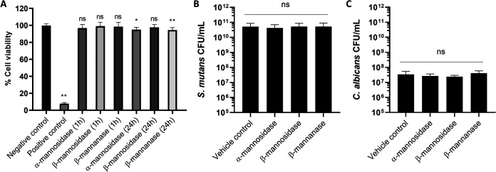
FIG 7.
Toxicity assay of MDEs on microbes and human gingival keratinocytes. (A) Normalized cell viability for HGKs after exposure to optimal units of MDEs for 1 h and 24 h, and CFU values for S. mutans (B) and C. albicans (C) after treatment with optimal units of MDEs for 5 min. No loss in HGK cell viability was observed for MDE treatments. Negative control and positive control represent vehicle control and 3% H2O2 control, respectively. There were no significant differences in microbial cell viability with or without MDE treatment. Statistics: one-way ANOVA with P < 0.01; post hoc: ** represents P < 0.0001 against vehicle control using Dunnett’s method (n ≥ 3); ns, not significant.

