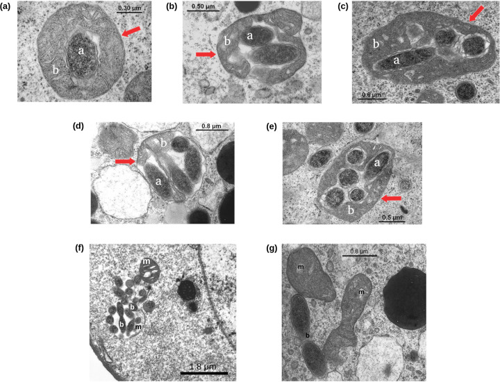
FIG 1.
Transmission electron microscopy observation of “Ca. Midichloria mitochondrii” bacteria in Ixodes ricinus oocytes. TEM images of mitochondria of Ixodes ricinus oocytes colonized by at least one (a), two (b), three (c), four (d), and five (e) “Ca. Midichloria mitochondrii” cells. In each photo, the red arrow indicates the mitochondrial membrane, the letter “a” indicates an intramitochondrial M. mitochondrii cell, and the letter “b” indicates the mitochondrial matrix. “Ca. Midichloria mitochondrii” cells possibly in replication within a mitochondrion (f) and in the cytoplasm (g); b, bacterium; m, mitochondrion.

