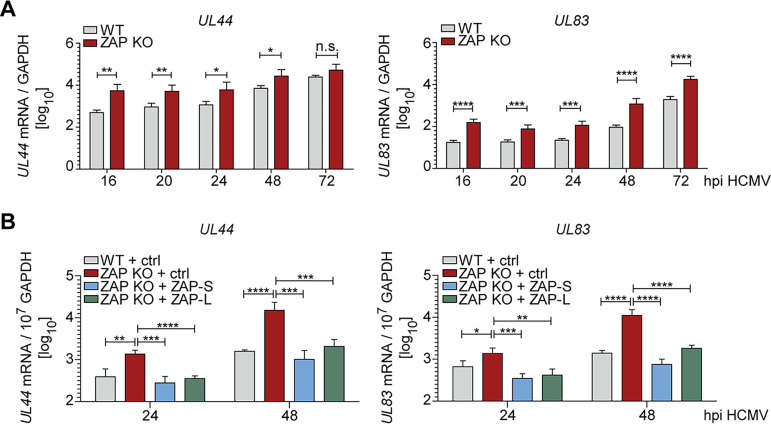
FIG 4.
ZAP-S and ZAP-L negatively affect early and late HCMV transcripts. (A) WT or ZAP KO HFF-1 cells were infected by centrifugal enhancement with HCMV (MOI 0.1). Total RNA was extracted at the indicated time points postinfection, and mRNA levels of HCMV UL44 and UL83 were measured by qRT-PCR. Viral mRNA relative expression (log10) normalized to GAPDH is displayed as bar plots showing the mean ± S.D. of three independent experiments performed with experimental duplicates. Experiments were performed in three independent ZAP KO cell lines, and results were combined. (B) ZAP KO HFF-1 stably expressing either ZAP-S or ZAP-L or transduced with empty vector control, and WT cells expressing empty vector, were infected by centrifugal enhancement with HCMV (MOI 0.1) and qRT-PCR performed as described in panel A. Viral mRNA relative expression (log10) normalized to GAPDH is displayed as bar plots showing the mean ± S.D. of two independent experiments performed with experimental duplicates. hpi, hours postinfection. Significant changes were calculated using unpaired two-sided Student’s t tests; n.s. not significant; *, P < 0.05; **, P < 0.01; ***, P < 0.001; ****, P < 0.0001.

