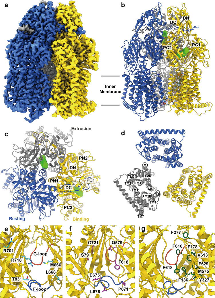
FIG 1.
Cryo-EM structure of apo-AdeJ. (a) Side view of the sharpened cryo-EM map of apo-AdeJ (viewed in the membrane plane). (b to d) Ribbon diagram of the structure of the side view (viewed in the membrane plane), top view (viewed from the extracellular space), and bottom view (viewed from the cytoplasm) of the apo-AdeJ trimer. In panels a to d, the resting, binding, and extrusion state protomers are colored royal blue, gold, and gray, respectively. The binding and extrusion channels are colored green. (e) Entrance drug-binding site. (f) Proximal drug-binding site. (g) Distal drug-binding site. In panels e to f, residues that are predicted to be important for selectivity are shown as sticks. The F-loop and G-loop are colored royal blue and tomato, respectively.

