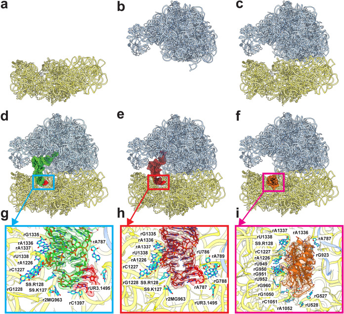
FIG 3.
Cryo-EM structures of the A. baumannii ribosome bound with Era. (a to f) Structures of the A. baumannii 30S, 50S, empty 70S, P-site tRNA 70S, E-site tRNA 70S, and hpf-bound 70S. All of these ribosomes are bound with Era. In panels d to f, the P-site tRNA and E-site tRNA are colored green and brown, respectively. The mRNA fragment is in red. The bound hpf is colored orange. (g) P-site tRNA-binding site. The cryo-EM density corresponding to bound tRNA is colored green. The density corresponding to the mRNA fragment is colored red. Nucleotides that are involved in tRNA binding are shown as cyan sticks. (h) E-site tRNA-binding site. The cryo-EM density corresponding to bound tRNA is colored brown. The density corresponding to the mRNA fragment is colored red. Nucleotides that are involved in tRNA binding are shown as cyan sticks. (i) hpf-binding site. The cryo-EM density corresponding to bound hpf is colored orange. Nucleotides that are involved in hpf binding are shown as cyan sticks.

