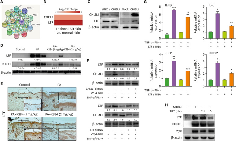
Figure 4. K284 reduces LTF expression. (A) Protein-association network analysis of Chi3L1 (Human). (B) The changes of CHI3L1-related proteins in the patients of AD obtained from ArrayExpress. (C) HaCaT cells were transfected with CHI3L1 plasmid vector or siRNA (20 nM) for 24 h. The levels of CHI3L1 and LTF were measured by Western blot analysis. (D) The protein expression levels of CHI3L1 and LTF in PA-treated skin tissues were measured by Western blot. (E) Expression of LTF in in PA-treated skin tissues analyzed by IHC. Scale bar=50 μm. (F) HaCaT cells were transfected with CHI3L1 of LTF siRNA for 24 h. Cell were pre-treated with K284 for 2 h. Then, cells were treated with TNF-α and IFN-γ (20 ng/ml) 4 h. The levels of CHI3L1 and LTF were determined by Western blot analysis. (G) HaCaT cells were transfected with LTF siRNA (20 nM). After 24 h, cells were treated with TNF-α and IFN-γ combination for 4 h. The mRNA expression of IL-1β, IL-6, TSLP and CCL22 was determined by qPCR (n=3).
#Control vs. TNF-α+IFN-γ and *TNF-α+IFN-γ vs. TNF-α+IFN-γ with LTF siRNA. *p<0.05, **p<0.01 and ***p<0.001.

