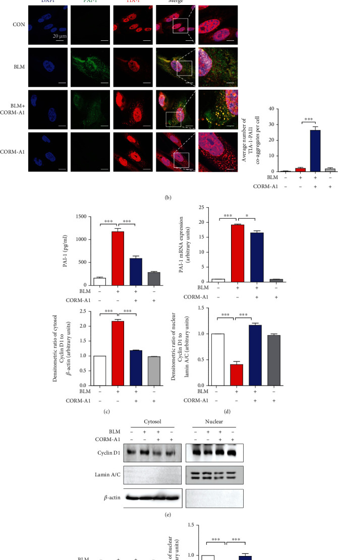
Figure 4.

CO inhibits secretion of PAI-1 in senescent cells via SG formation. (a) WI-38 cells were treated with CORM-A1 in various concentrations (0, 20, 40, and 80 μM) and iCORM-A1 (40 μM) for 6 h. As a positive control, WI-38 cells were treated with 200 nM thapsigargin (Tg) for 45 min, and then, immunofluorescence assay was performed to detect the formation of SGs by visualizing the colocalization of TIA-1(red) and G3BP1 (green). (b) WI-38 cells were pretreated with CORM-A1 (40 μM) for 6 h prior to the administration of bleomycin (25 μg/ml) for 96 h. After a 4-day treatment, immunofluorescence assay was performed to detect the TIA-1 (red) and PAI-1 (green) coaggregates. The secretion of PAI-1 was assessed by ELISA (c), and the mRNA expression of PAI-1 was assessed by qRT-PCR (d). (e) Nuclear translocation of cyclin D1 was measured by Western blot assay in cytosol and nuclear fractions. (f) The phosphorylated and total forms of Rb were analyzed by Western blot assay. The secretion of SASP, including IL-6 (g), TNF-α (h), and IL-1β (i), was measured by ELISA. Quantitative data are expressed as the means ± SD (n = 3 determined in three independent experiments). ∗∗p < 0.01 and ∗∗∗p < 0.001. ns: not significant.
