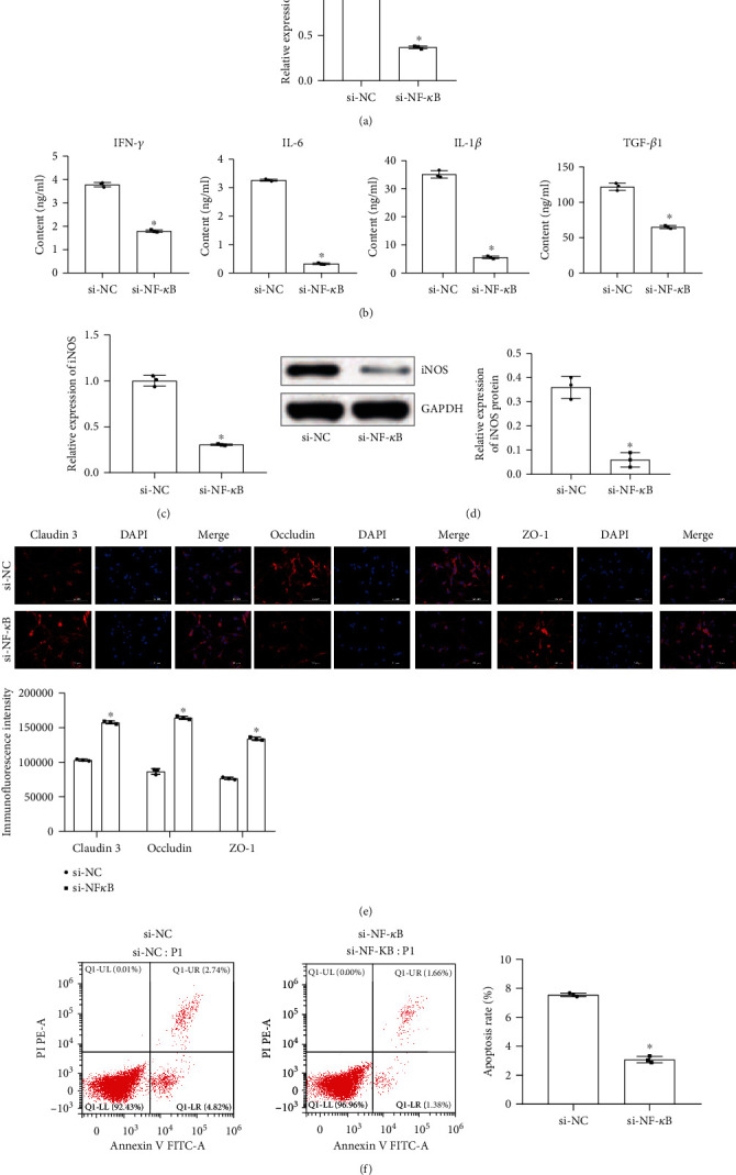
Figure 4.

Astrocytes were protected by inhibition of NF-κB. (a) qRT-PCR tested the expression of NF-κB. (b) ELISA was used to measure the content of IFN-γ, IL-6, IL-1β, and TGF-β1 in astrocytes. (c) qRT-PCR detected the expression of iNOS. (d) Western blot measured the protein expression of iNOS. (e) Fluorescence intensity of claudin 3, occludin, and ZO-1 was detected by immunofluorescence (scale bar, 100 μm). (f) Flow cytometry was used to measure the apoptosis rate of astrocytes. ∗Compared with the si-NC group, P < 0.05. The above experimental results were all measurement data. Data are expressed as the mean ± SD. The unpaired t-test was used between the two groups.
