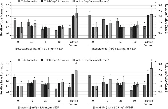
Fig 8. Anti-angiogenic compounds can abolish tube formation without induction of cell death.
HUVECs were seeded on a confluent layer of NHDFs on day 5 and cultured with 3.75 ng/ml VEGF-A165 (VEGF) from day 7 ± increasing concentrations of the anti-angiogenic compounds bevacizumab (0.01, 1 and 10 μg/ml), regorafenib (10, 50 and 100 nM), sorafenib (10, 25 and 50 nM), and sunitinib (10, 25 and 50 nM). At the end of the experiment (day 11), the co-cultures were fixed and stained for the endothelial marker PECAM-1 and the apoptotic marker active caspase-3 (Casp-3) to reveal tubes and apoptotic cells, respectively. Left axis: Relative tube formation. Tube formation is expressed relative to the untreated control. Right axis: Relative active caps-3. Active casp-3 is expressed relative to the VEGF treated sample. Active casp-3 masked PECAM-1: Active casp-3 in areas covered with HUVEC cells. The positive control was treated with 1 μM staurosporine for 6h prior fixation. Tubes were quantified with the 2cureX tube and casp-3 algorithm. Two individual experiments were performed, and representative results are shown. Data are the average of triplicate samples ± SD. *, †, # p<0.05 for anti-angiogenic treated vs. VEGF treated samples with respect to tube formation, total casp-3 activation in the sample and active casp-3 masked PECAM-1, respectively.

