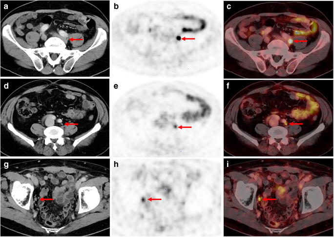
Fig. 3.
Examples of three different patients presenting with different tracer uptake intensities in histologically confirmed lymph node metastasis (LNM) despite comparable size of LN and clinical parameters. A-C: [68Ga]Ga-PSMA-11 PET/CT imaging of a 66-year old patient with recurrent PC (GSC 8; PSA level at PET examination 1.1 ng/ml). The patient presented with a correctly classified lymph node metastasis (LNM) behind the left common iliac artery with an intense, focal uptake on [68Ga]Ga-PSMA-11 PET (B, red arrow) and fused [68Ga]Ga-PSMA-11 PET/CT (C, red arrow). SUVmax of the LNM was 13.6. In the corresponding CT, only a small unsuspicious LN with a maximum diameter of 5 mm could be found (A, red arrow). D-F: 71-year old patient with PSA failure after radical prostatectomy (GSC 8; PSA level at PET examination 1.6 ng/ml) and a correctly classified LNM by [68Ga]Ga-PSMA-11 PET imaging: a morphologically completely unobtrusive lymph node is visible behind the left common iliac artery (axial diameter 5 mm) on sole CT imaging (A, red arrow) that shows intense, focal and thus suspicious tracer uptake on [68Ga]Ga-PSMA-11 PET (B, red arrow) and PET/CT fusion imaging (C, red arrow). SUV max of LNM was 4.1. G-I: 76-year old patient with biochemical recurrent PC (PSA value 0.77 mg/ml) after radical prostatectomy (GSC 9) presenting with a PSMA-positive LNM in the right obturator fossa (A and B, red arrow). Corresponding CT shows a slightly enlarged lymph node with an axial diameter of 10 mm (A, red arrow). SUV max of LNM was 5.1

